|
We recently reported that humans have a regulatory deletion downstream of the androgen receptor (AR) gene (McLean et al. (2011) Nature 471:216). This deletion removes a highly-conserved regulatory element from the AR gene that is normally expressed in small keratinized spines that form on the surface of the developing penis. Keratinized penile spines are found in many species, including both mice and chimps. Since formation of these spines is known to be dependent on androgen signaling, we proposed that the regulatory change in the human AR gene may explain the loss of similar penile spines in the human lineage (McLean et al. (2011)).
After the study was published, several people have written us to ask about the relationship between "penile spines" in mice and chimps and structures called "pearly penile papules" (PPP) in humans (also known as Hirsuties papillaris genitalis or Hirsuties coronae glandis). PPP are small projections, 1 to 3 mm in diameter, that form in variable numbers around the glans of the penis in approximately 20% of human males (Glicksman and Freeman (1966) Arch. Dermatology 93: 56-59). Despite their superficial resemblence to penile spines in other animals, PPP look very different at the cellular level.
The accompanying figure compares histological sections through the penile spines of mice and chimps, and a penile papule in humans.
In mice and chimps, penile spines are small projections made from the local piling up of cell layers in the outermost keratinized epidermis of the skin (top row).

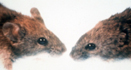
Mouse and chimp keratinized penile spines
Left panel: normal mouse penis, showing surface spine made of keratinized epithelium (Murakami (1987) J.Anat. 153:223). Middle panel: AR penile spine enhancer pattern, This is the regulatory element missing in humans (McLean (2011) Nature). Right panel: chimp penis, showing surface spine also made of keratinized epithelium (Hill (1946) ProcZoolSocLondon 116:129).

The different structure of a human penile papule
Unlike the keratinized spines shown above, the papules that form in some humans are outpockets of the entire skin surface, with a dense and vascularized connective tissue core making up the bulk of the projection. Although mouse and chimp penile spines are closely associated with neural sensory receptors, human PPP are not enriched in neural structures (see Glicksman and Freeman (1966) Arch Derm 93: 56-59).
In contrast, a PPP in humans consists of a larger out-pocketing of the entire skin, without local layering of the keratinized epidermis to make hook-like projections, and with the bulk of the overall structure consisting of a dense, well vascularized core of connective tissue cells, rather than epithelial cells (bottom row).
In addition, while mouse and chimp penile spines are often closely associated with neural sensory structures, human PPP do not appear to be enriched for neural cells (Glicksman and Freeman (1966) Arch. Dermatology 93: 56-59).
PPP are an interesting morphological variant in humans. However PPP are structurally distinct from the typical penile spines found in mice and chimps, and likely form by very different, still poorly understood mechanisms. Finally, we also note that the variable presence or absence of pearly penile papules in human males has no apparent relationship to the presence or absence of the penile spine regulatory sequence we have described in the androgen receptor gene. While PPP form in a significant fraction of humans, all humans tested appear to be missing the regulatory control sequence for keratinized penile spine formation described in the recent paper (McLean et al. (2011) Nature 471:216).
More information on research projects in mice, sticklebacks, human evolution, and human disease. |